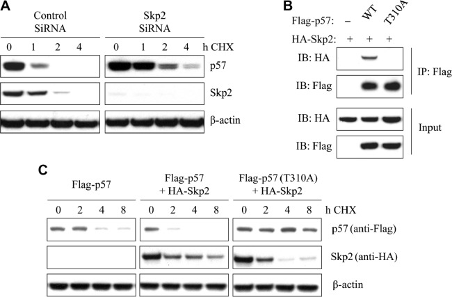
Figure 4.
Association of Skp2 with p57Kip2 degradation in breast cancer cell lines. (A) After transfection with Skp2 siRNA or control for 48 hours, T47D cells were incubated for the indicated times with CHX (20 μg/mL). Cell lysates were then subjected to immunoblot analysis with antibodys to p57Kip2 or Skp2. (B) Cells were transfected with HA-Skp2 and Flag-p57 (WT) or Flag-p57 (T310A) for 48 hours and then incubated with 10 μM MG132 for 4 hours. After that, cell extracts were immunoprecipitated with an antibody against Flag and analyzed by immunoblotting. (C) After transfection with HA-skp2 and Flag-p57 (WT) or Flag-p57 (T310A) for 48 hours, cells were incubated for the indicated times with CHX (20 μg/mL). Cell lysates were then used for immunoblot analysis with antibodies to Flag or HA. β-Actin was used as a loading control.

