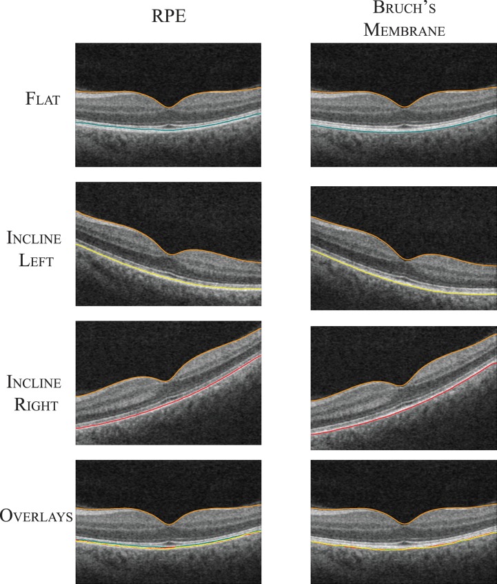
Figure 6.
A central B-scan from an example on-axis and two off-axis scans obtained from a normal subject. The segmented RPE and Bruch's membrane have been overlaid on the B-scan. The surfaces were transformed into the on-axis scan space using the ILM. The bottom row shows the segmented surfaces overlaid on the on-axis scan.

