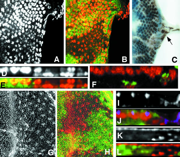
Figure 1.
Mbs mutant photoreceptors move basally and into the optic stalk. (A-C) Eye discs with MbsT666 clones stained with anti-Elav to label photoreceptor nuclei (A, red in B, brown in C). Wild-type tissue is marked with arm-lacZ, stained with anti-β-galactosidase in green (B) or X-gal in blue (C). Posterior is to the right. Many mutant cells are lost from the apical layer, and some Elav-positive nuclei are present in the optic stalk (arrow in C). (D and E) show cross sections of the discs in A and B, with apical up. Wild-type nuclei are close to the apical surface, but many mutant nuclei are found close to the basal surface. (F) Cross-section of an eye disc with MbsT666 mutant cells positively marked with CD8-GFP (green). Elav is stained in red. The entire membrane of Mbs mutant cells seems to be located basally within the disc. (G and H) Eye discs with MbsT666 clones stained with anti-phosphotyrosine (G, red in H) and with anti-β-galactosidase, marking wild-type tissue, in green in H. Apical constriction in the furrow is slightly reduced in the mutant tissue, and no ectopic constriction is visible in posterior regions. (I-L) Cross-sections of discs with MbsT541 clones stained with anti-E-cadherin (I, blue in J), anti-Elav (red in J), anti-Patj (K, red in L), and anti-β-galactosidase to mark wild-type tissue (green in J and L). E-cadherin and Patj are still expressed in mutant tissue, but at a more basal level than in wild-type tissue; staining is apical to mislocalized nuclei (J).

