Figure 4.
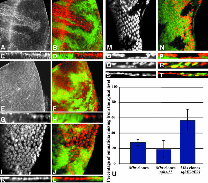
Sqh is the critical substrate for Mbs in eye development. (A-P) Eye discs containing Mbs clones, with wild-type tissue marked with anti-β-galactosidase staining in green (B, D, F, H, J, L, N, and P). (A-D) MbsT666 clones stained with an antibody to phospho-MRLC (A and C, red in B and D). (E-H) MbsT541 clones stained with an antibody to phospho-ERM (E and G red in F and H). Cross sections of the discs in A and B are shown in C and D, and cross sections of the discs in E and F are shown in G and H. p-Sqh is strongly up-regulated in Mbs clones, but the levels of p-Moe are unaffected, although p-Moe staining is normally at the apical surface and seems more basal in the clones. (I-L) show eye discs carrying the sqhA21 transgene and containing MbsT541 clones. Anti-Elav staining marks photoreceptor nuclei (I and K and red in J and L). (K and L) Cross sections of the disc in I and J. (M-P) Eye discs carrying the sqhE20E21 transgene and containing MbsT666 clones. Anti-Elav staining marks photoreceptor nuclei (M and O and red in N and P). (O and P) Cross sections of the disc in M and N. Nuclei are largely restored to the apical layer in Mbs clones by the presence of sqhA21 but are predominantly basal in the presence of sqhE20E21. (Q-T) Cross sections of eye discs containing MbsT666 clones, positively marked with GFP (green in R and T) and also expressing UAS-mycmoeT559A (Q and R) or UAS-mycmoeT559D (S and T) driven by tub-GAL4. Anti-Elav staining marks photoreceptor nuclei (Q and S and red in R and T). Neither transgene obviously alters the severity of the Mbs phenotype. (T) Quantification of the proportion of ommatidia completely missing from the apical layer in Mbs clones in a wild-type background or in a sqhA21 or sqhE20E21 background. p < 0.01 for a comparison of sqhA21 to wild type, and p < 0.001 for a comparison of sqhE20E21 to wild type.

