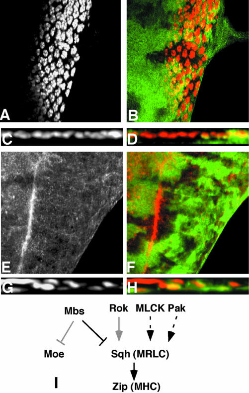
Figure 5.
Zip acts downstream of Mbs, but Rok is not the only upstream regulator of Sqh. (A-D) show eye discs heterozygous for zip1 and containing MbsT541 clones, with wild-type tissue marked with anti-β-galactosidase staining in green (B and D). Anti-Elav staining marks photoreceptor nuclei (A and C and red in B and D). (C and D) Cross sections of the disc in A and B. Removal of one copy of zip strongly suppresses the photoreceptor localization defect of Mbs mutant clones. (E and F) show an eye disc containing rok2 clones, with wild-type tissue marked with GFP (green in F). Phospho-MRLC is stained (E, red in F). p-Sqh is not obviously reduced in the absence of rok. (G and H) Cross sections of eye discs containing MbsT541 clones, positively marked with GFP (green in H) and also expressing UAS-RokCAT driven by tub-GAL4. Anti-Elav staining marks photoreceptor nuclei (G and red in H). Activated Rok does not strongly enhance the phenotype of loss of Mbs. (I) Functional relationships between components of the Mbs pathway. Arrows indicate activation and perpendicular lines inhibition. Black is used for interactions demonstrated to be important in the eye disc, gray for interactions that do not seem to occur in the eye disc, and dashed lines indicate possible interactions.

