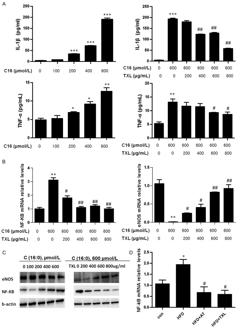
Figure 4.

TXL inhibited inflammation in C16-treated HCMECs. A. HCMECs were grown in six-well plates for 24 h, and then were stimulated with different doses of C16 for 12 h, or were pre-incubated with different doses of TXL before treating with 600 μmol/L of C16. The cultured medium of cells was harvested and the content of IL-1β and TNFαwas examined by ELISA. B. HCMECs were grown in six-well plates for 24 h, and then were pre-incubated with different doses of TXL before stimulating with 600 μmol/L of C16 for 24 h. the expression of eNOS and NF-κB was detected by real-time PCR. *P < 0.05, **P < 0.01 and ***P < 0.001 vs. C16-untreated group; #P < 0.05 and ##P < 0.01 vs. C16-treated group without TXL treatment. The bars represent the mean ± SEM. from 3 independent experiments. C. HCMECs were grown in six-well plates for 24 h, and then were stimulated with different doses of C16 for 12 h, or were pre-incubated with different doses of TXL before stimulating with 600 μmol/L of C16 for 24 h. The expression of eNOS and NF-κB was detected by Western blotting. D. ApoE-/- mice fed high-fat diet were orally administrated with TXL (0.75 g/kg/day). The expression of NF-κB in the thoracic artery was detected by real-time PCR. *P < 0.01 vs. control group; #P < 0.05 vs. HFD group (n = 5 in each group). The bars represent mean ± SEM.
