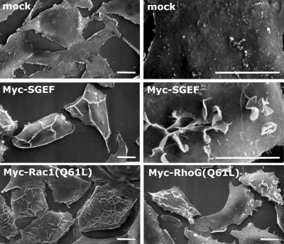
Figure 6.
Myc-SGEF expressing fibroblasts are morphologically similar to cells expressing activated RhoG. NIH3T3 cells were transiently transfected with expression plasmids for the indicated Myc-tagged protein, with mock cells receiving empty vector. Fibroblasts were then processed for scanning electron microscopy as described in MATERIALS AND METHODS. Myc-SGEF-induced dorsal ruffles were evident around cellular edges and across the dorsal surface of the fibroblast. Higher magnification revealed that the dorsal ruffles were highly connected with some degree of circular ruffling. Myc-Rac1(Q61L) expressing cells appeared flat with numerous dorsal ruffles that were shorter in stature than SGEF-induced ruffles. Cells expressing Myc-RhoG(Q61L) exhibited dorsal ruffles that were reminiscent of Myc-SGEF cells in both their connectivity and stature. Like Myc-SGEF cells, Myc-RhoG(Q61L) cells were less spread than Mock or Myc-Rac1(Q61L)-expressing cells. Bar, 10 μm.

