Figure 5.

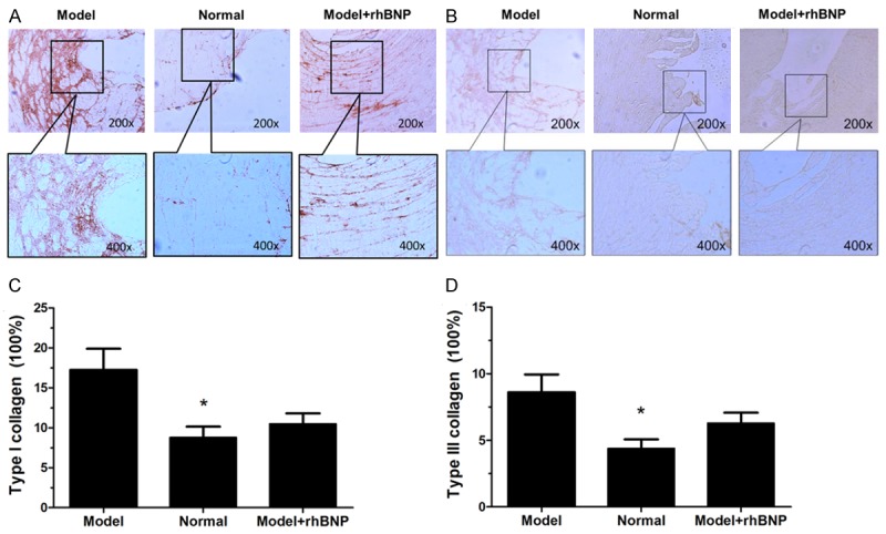
Immunohistochemical staining of type I and III collagen in post-MI tissues. A: Immunohistochemical staining of type I collagen in 3 tissues, C: The area percentage of type I collagen in 3 groups, B: Immunohistochemical staining of type III collagen in 3 tissues, D: The area percentage of type III collagen in 3 groups. *: the significance between Normal and Model group (P < 0.05).
