Figure 1.
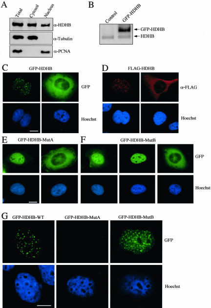
HDHB localizes in the nucleus or cytoplasm. (A) Cytoplasmic and nuclear extracts of U2OS cells were analyzed by denaturing gel electrophoresis and Western blotting with antibody against recombinant HDHB, α-tubulin, and PCNA. Immunoreactive proteins were detected by chemiluminescence. (B) GFP-HDHB transiently expressed in U2OS cells in contrast to endogenous HDHB. Control cells were transfected with pEGFP-C1 vector alone. (C) GFP-tagged HDHB, (D) FLAG-tagged HDHB, (E) Walker A (MutA), and (F) Walker B mutants (MutB) of GFP-HDHB transiently expressed in microinjected U2OS cells were visualized by fluorescence microscopy. Nuclei were stained with Hoechst dye. Bar, 10 μm. (G) U2OS cells transiently expressing GFP-HDHB wt, MutA, and MutB were extracted with 0.5% Triton X-100 before fixation and fluorescence microscopy. Nuclei were stained with Hoechst dye. Bar, 10 μm.

