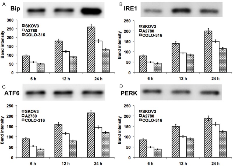
Figure 2.

Represent the western blot analysis of UPR related proteins Bip (A), IRE1 (B), ATF6 (C) and PERK (D) in DIM exposed ovarian cancer cells (SKOV-3, A2780 and COLO-316) for 24 hours. The represented blot pictures are correspondent to SKOV-3 cells. The graphical representation shows the quantifications of band intensity from the blots correspondent to all three ovarian cancer cell types.
