
Figure 1.
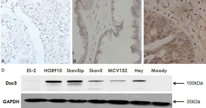
A-C. Expression of Dsc3 in different ovarian tissues determined by immunohistochemical staining (SP method). A. Benign ovarian cyst tissues; B. Borderline ovarian tumor tissues; C. Ovarian cancer tissues. Original magnification: ×100. D. Dsc3 expression was higher in the ovarian cancer cell lines HO8910, Skov3ip, Skov3, Hey and the borderline ovarian cystadenoma cell line MCV152 than the immortalized epithelial ovarian cells Moody.