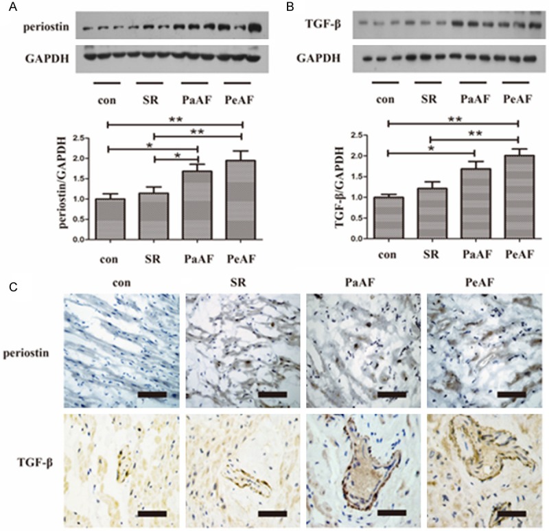
Figure 1.

Expression of periostin and TGF-β in RAAs. A, B. Representative western blot of periostin and TGF-β in RAAs. *P<0.05 between groups, **P<0.01 between groups. C. Representative immunohistochemical staining of periostin and TGF-β in different groups. Bar =50 μm. con group, n=10; SR group, n=20; PaAF group, n=15; PeAF group, n=25. SR, sinus rhythm; PaAF, paroxysmal atrial fibrillation; PeAF, persistent atrial fibrillation; TGF-β, transforming growth factor-β; RAAs, right atrial appendages.
