Figure 7.
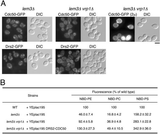
Localization of GFP-tagged Drs2 and Cdc50 proteins and the accumulation of NBD-labeled phospholipids in lem3Δ and lem3Δ vrp1Δ cells. (A) Localization of Cdc50p-GFP and Drs2p-GFP in lem3Δ and lem3Δ vrp1Δ cells. The lem3Δ (left column) and lem3Δ vrp1Δ (middle column) cells expressing either CDC50-GFP or DRS2-GFP were grown to early to mid logarithmic phase in YPDA medium at 25°C and observed immediately by microscopy. The lem3Δ vrp1Δ cells harboring both pKT1266 (YEplac181 CDC50-EGFP) and pKT1468 (YEplac195 DRS2) (right column) were grown to early-mid logarithmic phase in SD-Leu-Ura medium at 25°C and observed immediately by microscopy. GFP-tagged proteins were visualized using a GFP bandpass filter. For observation of each image, the same exposure and processing parameters were used. The strains used were as follows: YKT836 (Cdc50p-GFP in lem3Δ), YKT770 (Drs2p-GFP in lem3Δ), YKT805 (Cdc50p-GFP in lem3Δ vrp1Δ), YKT806 (Drs2p-GFP in lem3Δ vrp1Δ), and YKT843 (lem3Δ vrp1Δ) harboring pKT1266 and pKT1468. Bar, 5 μm. (B) Percentage of accumulation of NBD-labeled phospholipids of the deletion mutants relative to wild-type cells. pKT1472 (YEplac195 DRS2 CDC50) was introduced into YKT839 (lem3Δ vrp1Δ) strain. YEplac195 was introduced into YKT39 (WT), YKT496 (lem3Δ), and YKT839 (lem3Δ vrp1Δ) strains. Cells carrying the appropriate plasmids were grown to early-mid logarithmic phase in SD-Ura medium at 25°C and labeled with either NBD-PE, -PC, or -PS for 30 min at 25°C. The percentage of average accumulation for the deletion mutants relative to wild-type cell is presented with ±SD of seven independent experiments.

