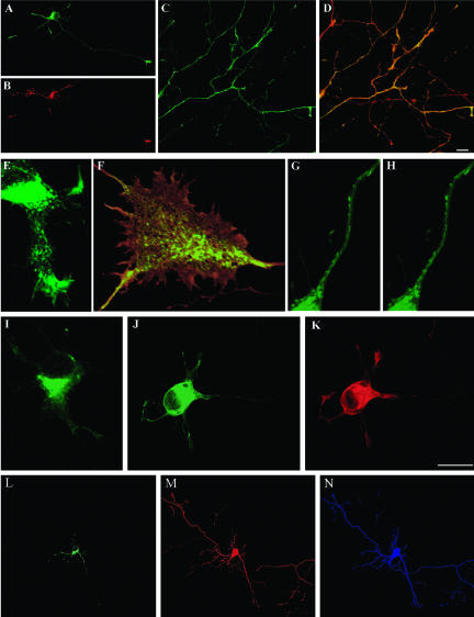
Figure 10.
LIMK1 regulation of the Golgi-derived export of NCAM-containing vesicles. (A and B) Double fluorescence micrographs showing the distribution of NCAM-GFP (A) and endogenous LIMK1 (B) in a cultured hippocampal pyramidal neuron. For this experiment, the cells were fixed 12 h after transfection and stained with the rabbit polyclonal antibody against LIMK1 used at a dilution of 1/1000. Note that the most intense NCAMGFP labeling is found within the cell body and at neuritic tips; light labeling is detected within minor neurites and along the axon (arrows) (C) Fluorescence micrograph showing the presence of NCAM-GFP along the axon and growth cones of a neuron cotransfected with HA-tagged wt-LIMK1. Note the intense NCAM-GFP labeling of the axon and growth cones. (D) Corresponding overlay image showing the distribution of NCAM-GFP (green) and HA-tagged wt-LIMK1 (red). The cells also were analyzed 12 h after transfection. (E) High-power confocal micrograph showing the presence of numerous NCAMGFP-containing vesicles within a neurite of a cell cotransfected with HA-tagged wt-LIMK1. (F) Abundant NCAM-GFP-containing vesicles (green) also are detected within growth cone-like structures (waves) located along the axonal shaft of a neuron cotransfected with HA-tagged wt-LIMK1 (red). (G and H) Series of confocal images showing the presence of numerous NCAM-GFP-containing vesicles in the axon of a neuron coexpressing HA-tagged Δ-PDZ LIMK1. (I and J) Immunofluorescence micrographs showing the distribution of NCAM-GFP in neurons cotransfected with HA-tagged LIMK1-kd. The cell shown in I was transfected with 2 μg/ml NCAM-GFP, whereas that shown in J with 4 μg of NCAM-GFP. Note that in both cases most of the labeling is localized to the cell body and absent from neurites and their tips. (K) Fluorescence micrograph showing the corresponding labeling of HA-tagged LIMK1-kd for the cell shown in J. Note that labeling is not only found in the cell body but also at neuritic tips. (L and N) Confocal images showing the distribution of NCAM-GFP (L) in a hippocampal neuron cotransfected with FLAG-tagged S3A cofilin (M) and HA-tagged wt-LIMK1 (N). Note that in this triple-transfected cell most of the NCAM-GFP labeling is found within the cell body. No labeling is detected in the middle or distal parts of neuritic processes, including growth cones. For all these experiments each LIMK1 cDNA was used at a concentration of 2 μg/ml. Bars, 10 μm (A-D, L and M) and 10 μm (E-K).

