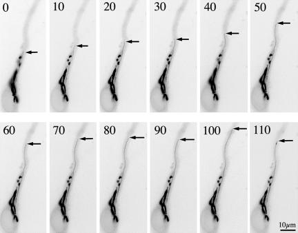
Figure 4.
Sequence of fluorescence images showing the dynamics of the Golgi apparatus from a neuron cotransfected with galactosyl-transferase T2-EYFP and FLAG-tagged S3A-cofilin. Note the extended morphology of the Golgi apparatus and the presence of a long tubulo-vesicular process (arrows) emerging from the Golgi stack that elongates and persists for more than a minute; other labeled tubules are out of the plane of focus. Images were taken every 10 s. For this experiment, cells were visualized 12 h after transfection.

