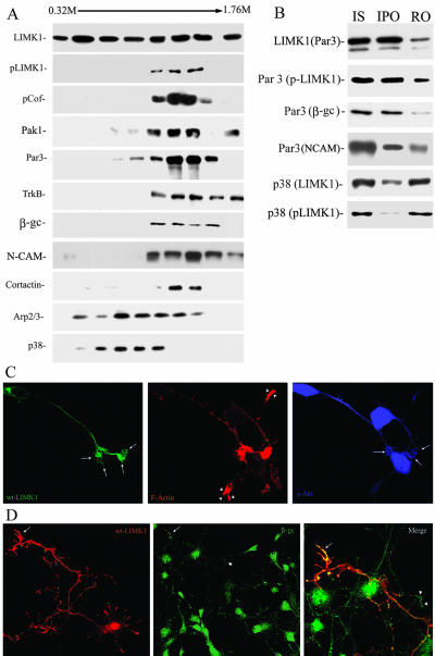
Figure 9.
Activated LIMK1 associates with Par3/Par6 containing vesicles. (A) Microsome fraction from developing rat cerebral cortex was fractionated by centrifugation across a continuous SDG and the same amount of protein (20 μg) from each fraction was applied to SDS-PAGE, stained with Coomassie Blue, or transferred to polyvinylidene difluoride membranes and analyzed by Western blot with antibodies against LIMK1, p-LIMK1, p-cofilin, Par3, TrkB, β-gc, NCAM, cortactin, Arp2, and synaptophysin (p38). Note the strong colocalization of p-LIMK1, p-cofilin, PAK1, Par3, TrkB, β-gc, and NCAM. (B) LIMK1 (Par3) Immunoisolation of Par3-containing organelles with anti-LIMK1 (dilution 1/50) from a microsomal fraction extending from 0.92 to 1.6 M sucrose; Par3 (LIMK1/p-LIMK1/β-gc/NCAM) Immunoisolation of p-LIMK1-, β-gc-, and NCAM-containing organelles with anti-Par3 (dilution 1/50). Note that all proteins are quantitatively recovered in the immunoprecipitated organelle fraction (IPO). Immunoisolation of LIMK1 and p-LIMK1-containing organelles with anti-p38 (dilution 1/50). Note that only a small amount of p-LIMK1 is associated with the immunoprecipitated organelle fraction. (C) Confocal images showing the distribution of HA-tagged wt-LIMK1 (green), F-actin (red), and p-Akt in a transfected hippocampal pyramidal neuron. Note the accumulation and intense labeling of p-Akt in the axonal growth cone (arrows) of the transfected neuron when compared with equivalent ones of nontransfected cells (arrowheads). (D) Confocal images showing the distribution of HA-tagged wt-LIMK1 (red) and β-gc (green) in a transfected hippocampal pyramidal neuron. Note the accumulation and intense labeling of β-gc in the axonal growth cone (arrows) of the transfected neuron when compared with nontransfected cells (arrowheads). Bar, 10 μm.

