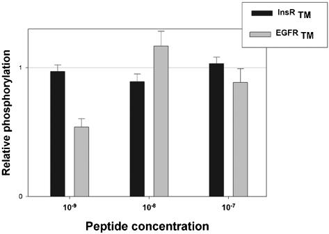
Figure 8.
Autophosphorylation of EGF receptors in A431 cells incubated with synthetic TM peptides maintained in solution in detergent. Peptides corresponding to the hydrophobic core of the TM domains of EGF (gray areas) and insulin (black areas) receptors, were chemically synthesized and purified, incorporated in detergent micelles, and administered to A431 cells by supplementation of the incubation medium for 4 h. After 5 min of incubation in the presence of 10-8 M EGF, cells were lysed, and equal amounts of protein were submitted to SDS-PAGE and Western blot with anti-EGFR and anti-phosphotyrosine antibodies. Densitometric analysis of the blots was performed, and results were normalized according to the amount of immunoreactive EGFR in each sample, and expressed as percentage of the observed autophosphorylation in the absence of peptides (taken as 100%). The figure represents the results (mean ± SEM) of four similar experiments.

