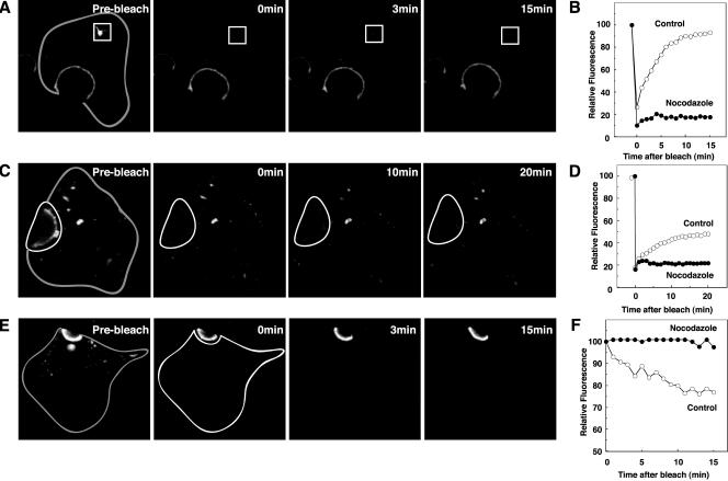
Figure 8.
Requirement of microtubules for constitutive BSEP cycling. After treatment with nocodazole (33 μM) for 60 min at 37°C, WIF-B9 cells infected with BSEP-YFP adenovirus were used for live cell imaging by confocal or conventional fluorescence microscopy. (A) Microtubule disruption perturbs recovery of globular structure BSEP pool. After treatment with nocodazole for 60 min at 37°C, globular structure pool enclosed by the white line was photobleached. Time in the subsequent two panels denotes minutes postbleach. Gray line shows basolateral membrane. (B) Quantitation of BSEP-YFP recovery in globular structure pool, which was photobleached as in A. Fluorescence intensity was determined as indicated. Solid circles denote nocodazole treatment and open circles are control values. (C) Microtubule disruption perturbs recovery of the canalicular BSEP pool. After treatment with nocodazole for 60 min at 37°C, canalicular area enclosed by the white line was photobleached. Time in the subsequent two panels denotes minutes postbleach. Gray line shows basolateral membrane. (D) Quantitation of BSEP-YFP recovery in the canalicular membrane pool which was photobleached as in C. Fluorescence intensity was determined as indicated. Solid circles indicate nocodazole treatment and open circles indicate control values. (E) Microtubule disruption perturbs BSEP-YFP loss in the canalicular pool after photobleaching the entire intracellular pool. Time in the subsequent two panels denotes minutes postbleach. Gray line shows basolateral membrane. (F) Quantitation of BSEP-YFP fluorescence change in the canalicular membrane pool. The entire cell except the canalicular membrane region was photobleached as in E. Solid circles denote nocodazole treatment and open circles are control values.

