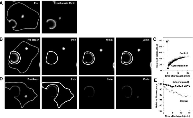
Figure 9.
Requirement of actin for constitutive BSEP cycling. After treatment with cytocholasin D (1 μg/ml) for 60 min at 37°C, WIF-B9 cells infected with BSEP-YFP adenovirus were used for live cell imaging by confocal microscopy at 37°C. (A) WIF-B9 cells infected with BSEP-YFP adenovirus were imaged pre- and postcytocholasin D treatment. Cytocholasin D treatment increased the canalicular BSEP pool. Gray line shows basolateral membrane. (B) Cytocholasin D treatment does not perturb BSEP recovery in the canalicular pool. After treatment with cytocholasin D for 60 min at 37°C, the canalicular region (white line) was photobleached. Time in the second two panels denotes minutes postbleach. Gray line shows basolateral membrane. (C) Quantitation of BSEP-YFP recovery in the canalicular membrane pool. The canalicular pool was photobleached as in C. Fluorescence intensity was determined as indicated. Solid squares denote cytocholasin D treatment and open circles denote control values. (D) Cytocholasin D perturbs BSEP retrieval from the canalicular pool. After treatment with cytocholasin D for 60 min at 37°C, the entire cell except the canalicular region was photobleached. Time in the second two panels denotes minutes postbleach. Gray line shows basolateral membrane. (E) Quantitation of BSEP-YFP fluorescence change in the canalicular membrane pool. The entire cell except canalicular region was photobleached as in D. Fluorescence intensity was determined as indicated. Solid squares denote cytocholasin D treatment and open circles denote control values.

