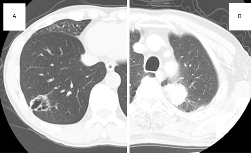
Figure 1.

Computed tomography findings. A. A chest computed tomography scan showing a cavitated mass, 33 × 30 × 28 mm, in the right lower lobe. The wall of the mass is relatively thicker than that of a typical benign cyst. B. A chest computed tomography scan showing a mass, 42 × 40 × 35 mm, with a serrated margin in the left upper lobe.
