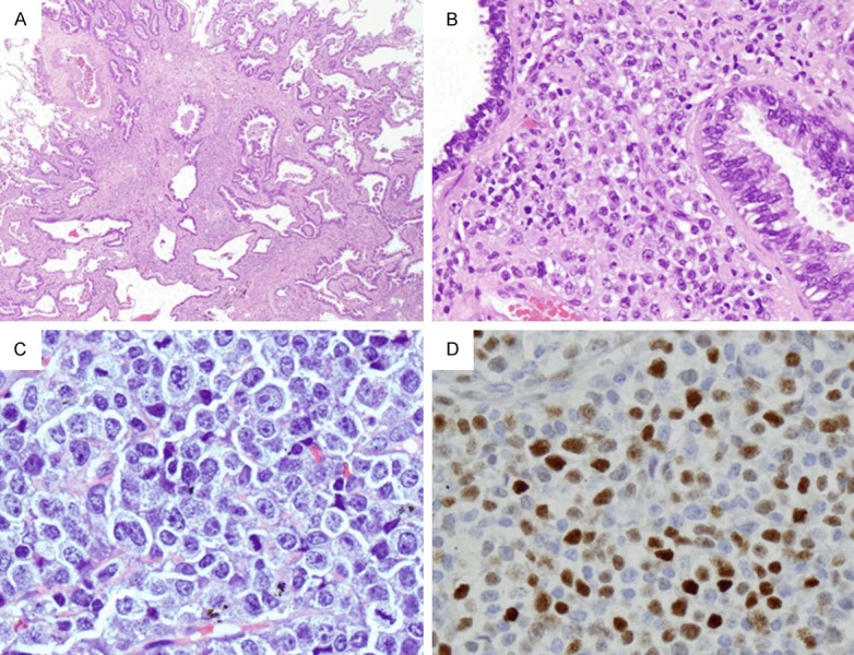
Figure 3.

(Case 3) Richter transformation in the lung and a lymph node from the neck. A and B. Show collision tumor of adenocarcinoma and diffuse large B-cell lymphoma in the lung parenchyma (A × 20; B × 200). Note the lymphoid infiltration is composed of large cells with prominent nucleoli. C. Shows lymph node with infiltration of similar atypical large lymphoid cells with slightly condensed chromatin and conspicuous nucleoli (× 400). D. Shows approximately 60% of these cells are positive for c-MYC (× 200).
