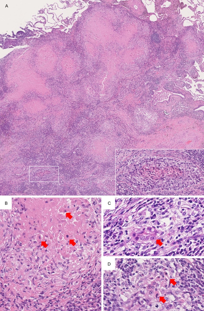
Figure 2.

Microscopic findings. A. Approximately 70% of the mass is composed of granulomatous inflammation accompanied by formation of prominent epithelioid cell granulomas with a central necrotic substance. In the remaining 30% of the mass, viable well-to-moderately differentiated squamous cell carcinoma is present (×20). Inset: Higher magnification of the boxed area shows a viable tumor nest with keratinization (×200). B. The necrotic substance inside the granulomas were consists of necrotic tumor cells that retained cellular shape and exhibited pyknotic nuclei (arrows) (×400). C. Tumor nest surrounded by macrophages and/or epithelioid histiocytes (arrow) (×400). D. Tumor nest with degeneration that showed remaining necrotic tumor cells with retained cellular shape and pyknotic nuclei (arrows) (×400).
