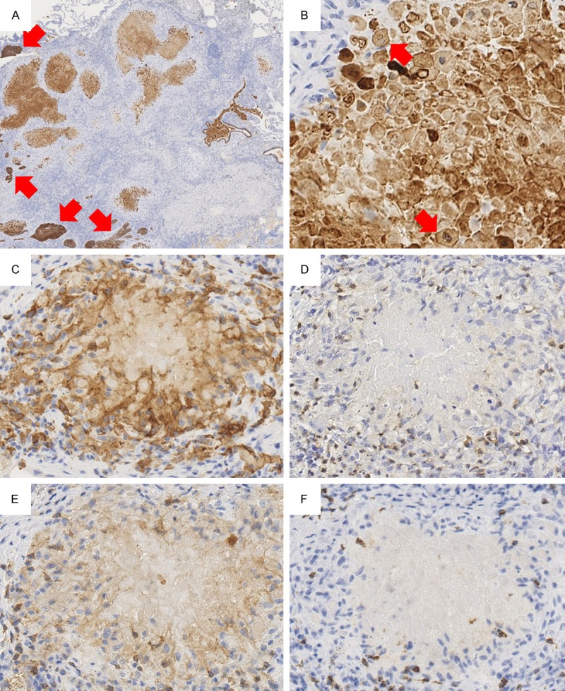
Figure 3.

Immunohistochemistry findings. (A) Viable tumor nests showing strong staining for CK5/6 (arrows). Necrotic substance showing moderate staining for CK5/6 (×20). (B) Necrotic substance inside a granuloma showing CK5/6-positive necrotic tumor cells that retained cellular shape and pyknotic nuclei (arrows) (×400). (C) Granulomas are composed of CD68-positive macrophages and/or epithelioid histiocytes (×400). (D) T lymphocytes expressing CD3 are abundant inside and outside the granulomas (×400). (E, F) Among T lymphocytes, CD4-expressing lymphocytes (E) are slightly more numerous than CD8-expressing lymphocytes (F) (×400).
