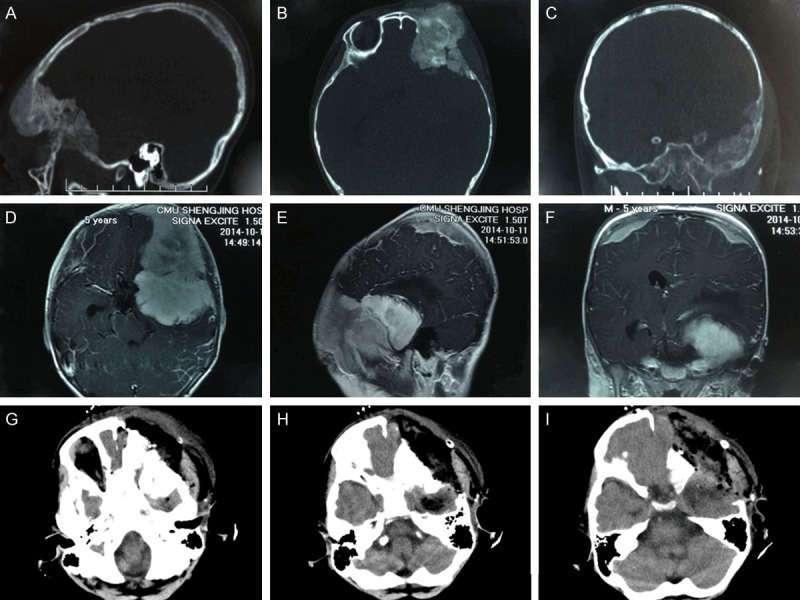
Figure 1.

A-C. Preoperative CT (sagittal, axial, and coronal scans, respectively) showing a hyperdense lesion in the greater sphenoid wing extending into the left orbital cavity, middle cranial fossa, and temporal fossa accompanied by destruction and thickening of biparietal bones. D-F. Preoperative MRI (axial, sagittal, and coronal scans, respectively) showing three enhanced masses in anterior and middle cranial fossa and biparietal bones. G-I. Postoperative axial CT scan showing that the mass in the middle cranial fossa was residual, and frontal and temporal bones invaded by the tumor were resected.
