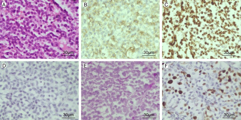
Figure 2.

(A) Hematoxylin and eosin-stained tumor specimen showing densely packed, small round cells with scanty clear cytoplasm and regular vesicular and hyper chromatic nuclei (B-D). Immunohistochemical analyses showing positive membranous staining for CD99 (B), positive staining for Vimentin (C) and negative for neuron-specific enolase (NSE) (D). (E) Periodic acid Schiff staining of the tissue showing the presence of glycogen granules. (F) Immunohistochemical staining for Ki-67. The tumor exhibited 40% Ki-67 labeling index.
