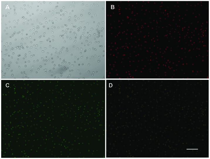
Figure 1.
Immunofluorescence identification and immunophenotype of bone marrow derived-EPCs. (A) Attached cells exhibited a spindle shaped, endothelial cell-like morphology. (B) Adherent cells 1,1-dioctadecyl-3,3,3,3-tetramethylindocarbocyanine (DiI)-labeled acetylated low-density lipoprotein uptake (red; excitation wave-length, 543 nm) and (C) lectin binding (green; excitation wave-length, 477 nm) were assessed under a fluorescence microscopy. (D) Double positive cells (yellow; overlay of B and C) were identified as differentiating EPCs. Magnification, ×200; scale bar, 100 μm. EPCs, endothelial progenitor cells.

