
Figure 6.
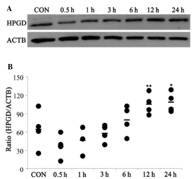
Expression of HPGD following hydroxychloroquine stimulation. The RA-FLS were serum-starved for 2 h and subsequently treated with hydroxychloroquine (10 µM) for 0.5, 1, 3, 6, 12 and 24 h. (A) The expression levels of HPGD in RA-FLS was determined by immunoblotting. ACTB was used as an internal control. (B) The immunoblotting was quantified and the data are expressed as the mean ± standard deviation (111.23±16.07, *P= 0.012; 106.99±16.82, **P=0.019, compared with the CON; n=4). CON, control; HPGD, 15-hydroxyprostaglandin dehydrogenase; ACTB, β-actin; RA-FLS, rheumatoid arthritis fibroblast-like synoviocytes.