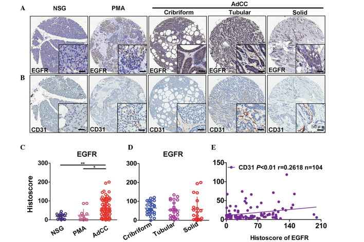
Figure 1.
Association between the expression of EGFR and CD31 in NSG, PMA and AdCC tissues. Representative immunohistochemical staining of (A) EGFR membranous expression and (B) CD31 membranous expression in human NSG, PMA and cribriform, tubular or solid type AdCC tissues. Scale bar=50 µm. Quantification of EGFR expression levels in (C) human NSG, PMA and AdCC tissues and (D) subtypes of AdCC using an AperioScanscope scanner and software. Data were analyzed by Graph Pad Prism 5 software. Data are presented as the mean ± standard error of the mean. *P<0.05, AdCC vs. PMA tissues; **P<0.01, AdCC vs. NSG tissues. (E) Correlation between EGFR and CD31 expression levels in human NSG, PMA and AdCC tissues (P<0.01, r=0.2618, n=104) using two-tailed Pearson's test. EGFR, epidermal growth factor receptor; NSG, normal salivary gland; PMA, polymorphism adenoma; AdCC, adenoid cystic carcinoma.

