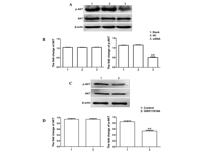
Figure 6.
Expression of Akt and phosphorylated (p)-Akt in the various groups. (A) Western blotting was used to analyze the protein expression of AKT and p-AKT in U937 human leukemia cells following transfection with small interfering (si)RNA-103. (B) Quantitative analysis of western blotting. **P<0.01, compared with the control. (C) Expression of AKT and p-AKT in U937 cells following treatment with GW311616A. (D) Quantitative analysis of western blotting. Data are presented as the mean ± standard deviation. NC, negative control.

