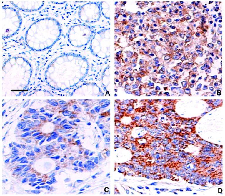
Figure 1.
Immunohistochemical staining for δ-catenin in normal colorectal tissues and colorectal cancer tissues. (A) Weak expression of δ-catenin was observed in the cytoplasm of normal colorectal gland epithelial cells. Scale bar, 50 µm. (B and C) Obviously enhanced expression of δ-catenin was observed in (B) the cytoplasm of poorly differentiated colorectal cancer cells and (C) highly differentiated colorectal cancer cells. (D) The expression of δ-catenin was also higher in lymph node metastases, compared to the corresponding primary tumor foci in C. Magnification, ×400.

