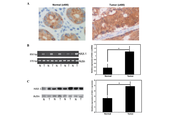
Figure 1.
Expression of HAX-1 in CRC tissue. (A) Representative immunohistochemical images of CRC tissue (magnification, x400), Left, normal tissue; right, tumor tissue). The expression levels of HAX-1 in CRC tissues were observed to be significantly higher than those in benign/adjacent noncancerous tissues. (B) mRNA expression levels of HAX-1 in colorectal cancer tissues (Left, reverse transcription-quantitative polymerase chain reaction; right, normalized with actin.) *P<0.05. Values are presented as the mean ± standard deviation. (C) Protein expression levels of HAX-1 in CRC tissues (Left, western blotting; right, normalized with acctin). *P<0.05. Values are presented as the mean ± standard deviation. HAX-1, hematopoietic cell specific protein 1-associated protein X-1; N, normal tissue; T, tumor tissue. CRC, colorectal cancer.

