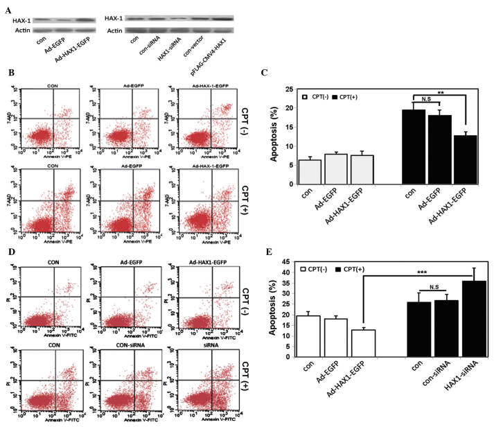
Figure 2.
Effects of the expression of HAX-1 on apoptosis. (A) SW480 human colorectal cancer cells were transfected with an HAX-1 overexpression vector (upper panel) or an HAX-1-siRNA vector (lower panel) for 48 h. The protein expression levels of HAX-1 were detected using western blotting. (B) Representative annexin V/7-AAD dot-plot distributions of SW480 cells treated without (upper panel) or with (lower panel) CPT in the different groups: Con, SW480 cells without infection; Ad-EGFP, SW480 cells infected with empty vector; Ad-HAX-1-EGFP, SW480 cells infected with Ad-HAX-1-EGFP vector. (C) Quantification of apoptosis. Experiments were repeated at least three times. Values represent the mean ± standard deviation (**P<0.01). (D) Representative annexin V/PI dot-plot distributions of transfected SW480 cells treated without (upper panel) or with (lower panel) CPT in the different groups: Con, SW480 cells without transfection; con-siRNA, SW480 cells transfected with control siRNA; siRNA, SW480 cells transfected with HAX-1-siRNA. (E) Quantification of apoptosis. Experiments were repeated at least three times. Values represent the mean ± standard deviation (***P<0.001). HAX-1, hematopoietic cell-specific protein 1-associated protein X-1; siRNA, small interfering RNA; CPT, camptothecin; siRNA, small interfering RNA; N.S, not significant.

