Figure 1.
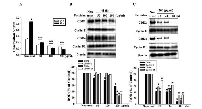
Fucoidan inhibits the proliferation of HT-29 cells. (A) HT-29 cell proliferation was measured using an MTT assay. Values are expressed as the mean ± standard error of the mean from three independent experiments (*P<0.05, **P<0.01, vs. 48 h fucoidan treatment). (B) HT-29 cells were incubated for 48 h with various concentrations of fucoidan (0, 50, 100 and 200 μg/ml) and the expression levels of CDK2, cyclin E, CDK4 and cyclin D1 were assessed by western blotting. (C) HT-29 cells were treated with fucoidan for different durations (0–48 h). The expression levels of CDK 2, cyclin E, CDK4 and cyclin D1 were assessed by western blotting. Values are expressed as the mean ± sandard error of the mean of four independent experiments for each condition, as determined from densitometry against to β-actin (*P<0.05, vs. non-treated cells). ROD, relative optical density.

