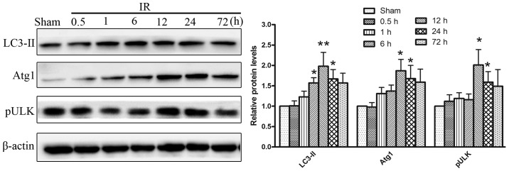
Figure 3.

Western blot analysis of autophagy-associated protein expression levels following cerebral IR injury. The levels of Atg1, pULK and LC3 in the ischemic cortex increased significantly between 1 and 24 h reperfusion, with a maximal induction at 12 h. *P<0.05 vs. sham group; **P<0.01 vs. sham group. IR, ischemia reperfusion.
