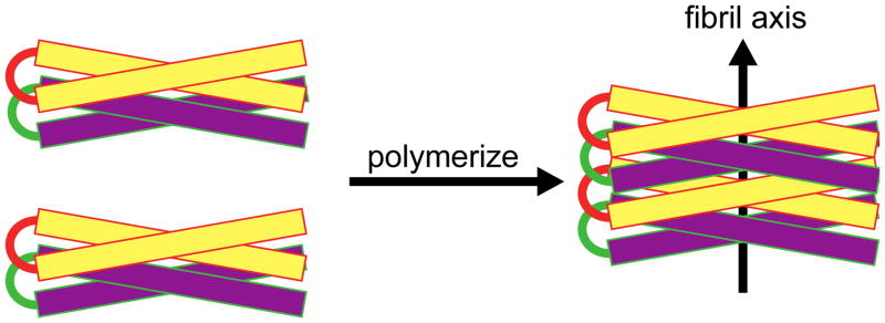
Figure 9.

Model of peptide fibrils. The α-helical axes are perpendicular to the long axis of the fibril. Helix-turn-helix dimers are shown stacking in a parallel manner to grow the individual fibril. For clarity, two colors (yellow and purple) are used to show the helix-turn-helix peptides stacking.
