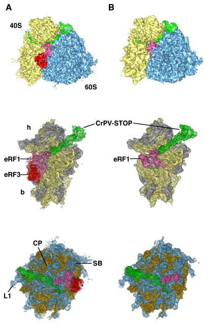
Figure 2. Cryo-EM maps of the 80S•CrPV-STOP•eRF1•eRF3•GMPPNP (A) and 80S•CrPV-STOP•eRF1 (B) termination complexes.
In vitro termination complexes were prepared on CrPV-STOP mRNA (green). Upper panel displays the 80S•CrPV-STOP•eRF1 eRF3•GMPPNP and the 80S•CrPV-STOP•eRF1 complexes with 40S subunit shown in yellow, 60S subunit in blue, eRF1 in hot pink and eRF3 in red. Middle and bottom panels display the corresponding 40S and 60S subunits with docked ligand models, respectively. Ribosomal RNA is shown in yellow or blue and ribosomal proteins in gray or orange for the 40S and 60S subunit, respectively. Landmarks of the 40S subunit are the head (h) and the body (b) domains. Landmarks of the 60S subunit: central protuberance (CP), the L1 stalk (L1) and the stalk base (SB).

