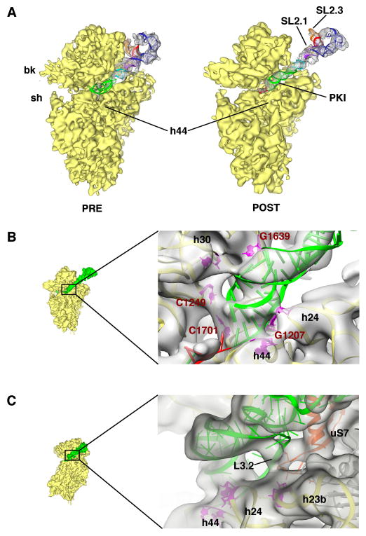
Figure 3. Post-translocational state of the CrPV IGR IRES and its interactions with the 40S subunit.
(A) Extracted densities for CrPV IRES and ribosomal 40S subunit are shown for the PRE- (left) (Schüler et al., 2006) and POST-translocational state from the 80S•CrPV-STOP•eRF1 complex (right). (B) Close-up view of the apical tip of PK I (green). Interactions to conserved nucleotides G1639, C1249, C1701 and G1207 are depicted (red) as well as interactions to h24 and h44. (C) Interactions of loop L3.2 of CrPV IRES (green) with the ribosomal protein uS7 and helix 23b.

