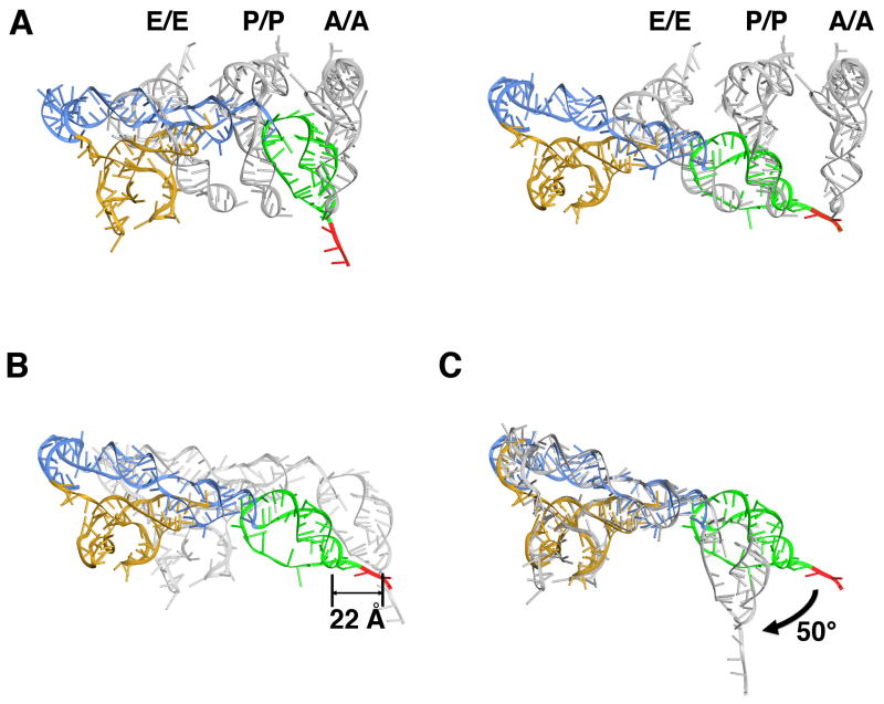
Figure 4. Comparison of the POST state IRES with tRNA positions and with PRE state IRES.
(A) Superposition of the PRE state IRES (left) (Fernández et al., 2014) and POST state IRES in the 80S•CrPV-STOP•eRF1 complex (right) with positions of canonical bound tRNAs (Budkevich et al., 2011) after 60S alignment. The CrPV IRES is colored according to the domain organization: domain 1 blue, domain 2 gold, domain 3 green. The first codon of the open reading frame is depicted in red. (B) Superposition of the PRE state IRES (gray) (Fernández et al., 2014) with the POST state IRES after corresponding 60S alignment. (C) Superposition of the PRE (gray) and POST state IRES after alignment of the corresponding ribosome-binding domains (domains 1 and 2).

