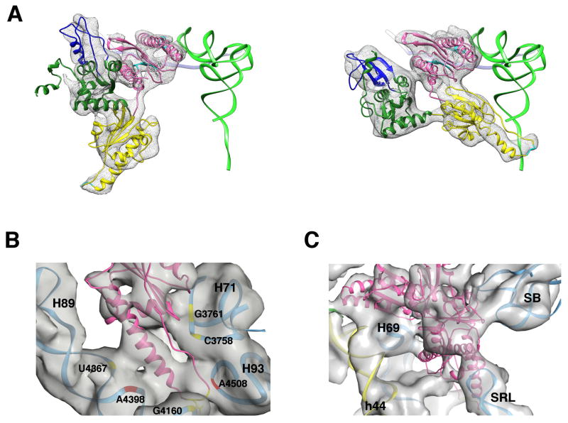
Figure 7. Ribosomal contacts of eRF1 in the 80S•CrPV-STOP•eRF1 complex.
(A) eRF1 conformation in the 80S•CrPV-STOP•eRF3•eRF1•GMPPNP (left) and the 80S•CrPV-STOP•eRF1 complex (right). As a marker, the canonical P-site bound tRNA is shown in green. eRF1 model is colored according to its domain organization: N domain in pink, M domain in yellow, C domain in green with the minidomain in blue. (B) The M domain of eRF1 is positioned in the PTC on the 60S subunit and forms conserved (red) as well as unique (yellow) ribosomal contacts. (C) In the 80S•eRF1 complex, the tip of the SRL interacts with the C domain of eRF1, which is analogous to the eukaryote-specific interaction involving the T-loop of A/T tRNA seen in the decoding complex (Budkevich et al., 2014).

