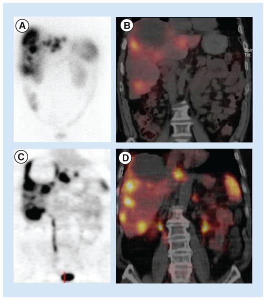
Figure 3. Comparison of (A) planar OctreoScan, (B) fused OctreoScan/SPECT/CT, (C) planar 68Ga-DOTATOC PET and (D) 68Ga-DOTATOC PET/CT in the same patient.
The patient has approximately 33% of their liver replaced by neuroendocrine tumor metastases. The images in (C) and (D) provide more precise delineation of lesions, compared with (A) and (B).

