
Figure 2.
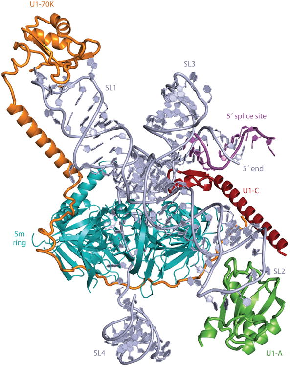
Overview of a model of the complete human U1 small nuclear ribonucleoprotein (snRNP) derived from X-ray crystal structures. Truncated stem loop 2 (SL2) was extended with an A-form RNA helix and, using the crystal structure of the U1A–RNA complex (64), was appended to the extended helix. The internal loop of SL2, consisting of four consecutive non-Watson-Crick base pairs, is in a position to interact with the Sm-B and Sm-D1 proteins. Closely matching images are found in the gallery of negatively stained images of U1 snRNP reported previously (251, 252).