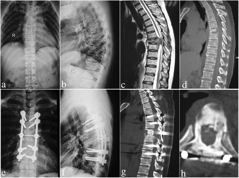
Fig. 3.

A 52-year-old female with T5/6 lesions was performed by posterior-only approach. a-d The pre-operative imaging data showed T5/6 vertebral bodies’ destructions with mild kyphosis deformity and spinal cord severely compressed. The postoperative anterior-posterior (e) and lateral X-ray (f) indicated that the kyphosis got obviously improved by posterior long-segment fixation. Sagittal and coronal CT-scan (g, h) showed satisfied allograft fusion without relapse of Pott’s disease at the 9 months of post-operation
