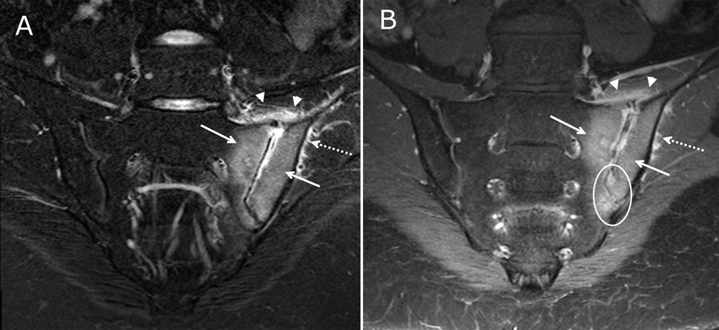
Figure 1. Active inflammatory lesions of the sacroiliac joints visible on both fluid-sensitive and post-contrast sequences.
17-year-old male with left lower back pain. (A) Coronal oblique STIR imaging of the sacrum demonstrates bone marrow edema within the inferior periarticular aspect of right iliac bone (solid arrow). There is a small amount of joint fluid within the inferior aspect of the joint (arrowhead). A normal hyperintense subchondral stripe is seen on the left in this skeletally immature child (dashed arrow). (B) Coronal oblique T1 weighted post contrast imaging of the sacrum demonstrates enhancing bone marrow edema within the inferior right iliac bone (solid arrow) and adjacent synovitis within the inferior, ventral aspect of the joint (arrowheads) with adjacent periarticular enhancement which is greater on the iliac side. There is a normal, mild enhancement of the left segmental sacral apophyses (dashed arrow).

