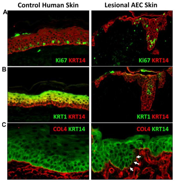
FIG. 2.
Cellular abnormalities observed in the skin of AEC patients. A: Proliferation, as shown here by Ki67 immunofluorescent staining (green) is restricted to the basal layer in normal human skin, but extends suprabasally in AEC patient skin. B: The marker for terminal differentiation, KRT1 (green/yellow), is induced in the first suprabasal layer in control human skin, but is delayed in its expression or absent from AEC patient skin. C: Immunofluorescent staining for the basement membrane component Collagen IV (COL4; red) shows uninterrupted staining along the basement membrane in control human skin, but is disrupted in AEC patient skin. Immunofluorescent staining for keratin 14 (KRT14) was used to highlight the epidermis in A (red), B (red), and C (green). Image reproduced from [Koster et al., 2009].

