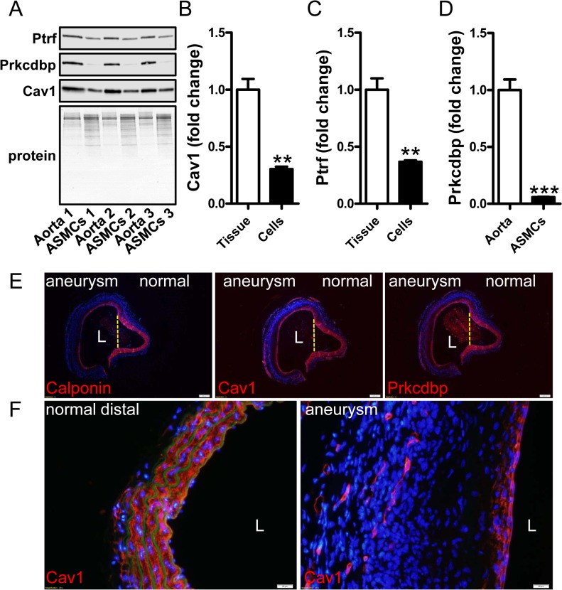
Fig 7. Caveolin-1 and cavin-1 conform to patterns of regulation established for classical smooth muscle differentiation markers.
Aortae from three different control mice were isolated. One piece was immediately frozen and the other was used for isolating smooth muscle cells which were subsequently cultured in vitro. (A) Western blots show higher expression of cavin-1 (Ptrf) and -3 (Prkcdbp) and caveolin-1 (Cav1) in whole aortae than in cultured cells from the same vessels. Panels B-D show summarized western blot data. Data are presented as means±SEM.**P<0.01, ***P<0.001. Panel E shows staining (in red) for calponin (left), caveolin-1 (middle) and cavin-3 (Prkcdbp, right) in consecutive sections from a saccular aortic aneurysm induced by AngII. This aneurysm had a diameter exceeding 2 mm and the lumen is indicated by an L. The aneurysm bulges to the left whereas the right side is normal. The partitions between normal and diseased areas are indicated by dotted yellow lines. A progressive reduction, from the normal to the dilated side, of medial staining for calponin, caveolin-1 and cavin-3 is evident, leaving only a thin red rim just below the endothelium at the aneurysmal site. This is shown at higher magnification in panel F, giving views of caveolin-1 staining in distal control aorta (left) and in the aneurysm (right). Caveolin-1-positive capillary structures are present in the adventitia of the aneurysm whereas medial staining is drastically reduced. Nuclei were stained with DAPI throughout. In panel F, autofluorescence (green) was used to visualize elastic lamellae showing loss of elastic fibers in the aneurysm to the right. Images are representative of three aneurysms and three control aortae. Scale bars (white) in E and F represent 200 and 20 μm, respectively.

