Figure 2.
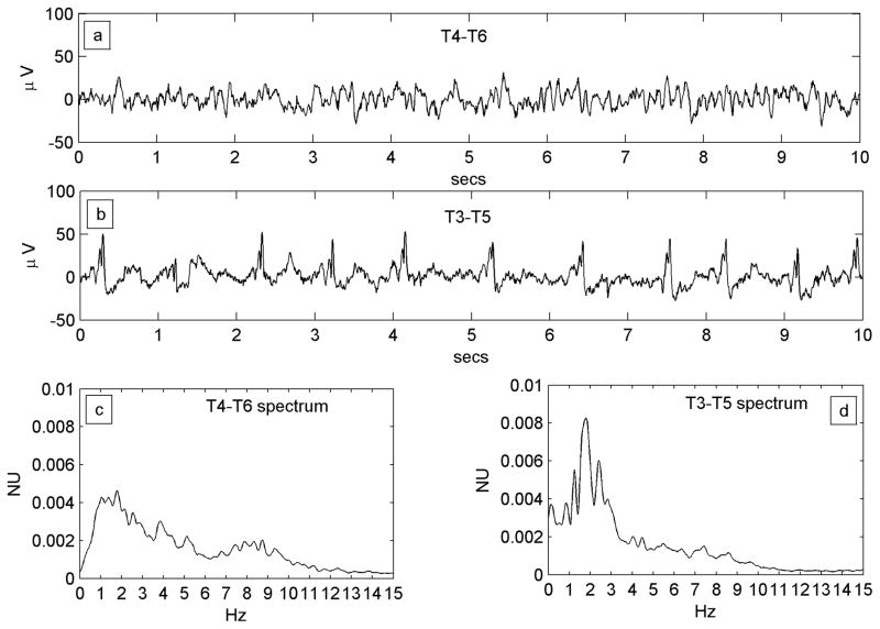
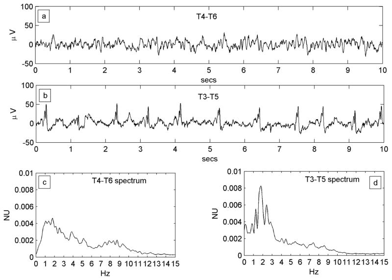
Figure 2A. A regular PLED in a patient with an acute left posterior quadrant lobar hemorrhage (Patient #1) illustrating condensation and low-pass filtering. a) A10-s EEG epoch from the normal right hemispheric mid-temporal derivation, showing a mix of polymorphic fast and slow rhythms and irregular larger amplitude transients b) A typical epoch of the homologous left mid-temporal chain showing PLEDs at frequency ~1 Hz. c) Power spectrum of the entire T4–T6 EEG epoch. The spectrum is wide-band with broad maxima in the δ and α Berger bands, the latter indicating a preserved posterior dominant rhythm. d) Power spectrum of the entire T3–T5 EEG epoch. Comparison of the spectra illustrates spectral condensation, with the broad δ band of the normal side appearing to coalesce into sharply defined peaks in the PLED spectrum. Indeed, the small local maximum on the normal side (at ~2 Hz) ‘grows’ in the same location into the dominant frequency component of the PLED. The two spectra also illustrate low-pass filtering, with the α-band frequencies on the normal side flattening out on the PLED side, in keeping with the visually-obvious absence of the α rhythm in the raw PLED time series.
Figure 2B. Example of the condensation-plus-harmonics transformation in Patient #7 with serial seizures and a previously resected brain tumor. a) Continuous slowing on the normal (but encephalopathic) left side. b) Right-sided ~1 Hz PLED pattern. c) Left sided power spectrum shows a relatively narrow base with significant power only below ~4 Hz in keeping with the slowing seen in the time series. A prominent peak at ~2 Hz is however present. d) The PLED spectrum shows more discrete and condensed appearance, with three main peaks: a fundamental (at ~1 Hz), a second harmonic (at ~2 Hz) that corresponds to the maximum peak of the normal side, and a third harmonic (at ~3 Hz). A further smaller peak at ~3.5 Hz appears, of uncertain origin.


