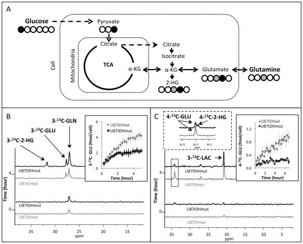
Figure 4. Glucose and glutamine flux to glutamate is reduced in U87 IDH1 mutant cells.
Schematic representation of 13C-labeling of glutamate and 2-HG derived from 1-13C-glucose and 3-13C-glutamine (A). Build-up of 3-13C-glutamate (3-13C-GLU) and 3-13C-2-HG following perfusion of U87IDHwt and U87IDHmut cells with 3-13C-glutamine (3-13C-GLN) with quantification of 3-13C-glutamate production shown in the inset (B). Build-up of 3-13C-lactate (3-13C-LAC), 4-13C-glutamate (4-13C-GLU) and a combined signal of 4-13C-glutamate and 4-13C-2-HG in U87IDHwt and U87IDHmut cells with quantification of the build-up of glutamate in right inset. Left inset illustrates data from cell extracts resolving 2-HG and glutamate (C).

