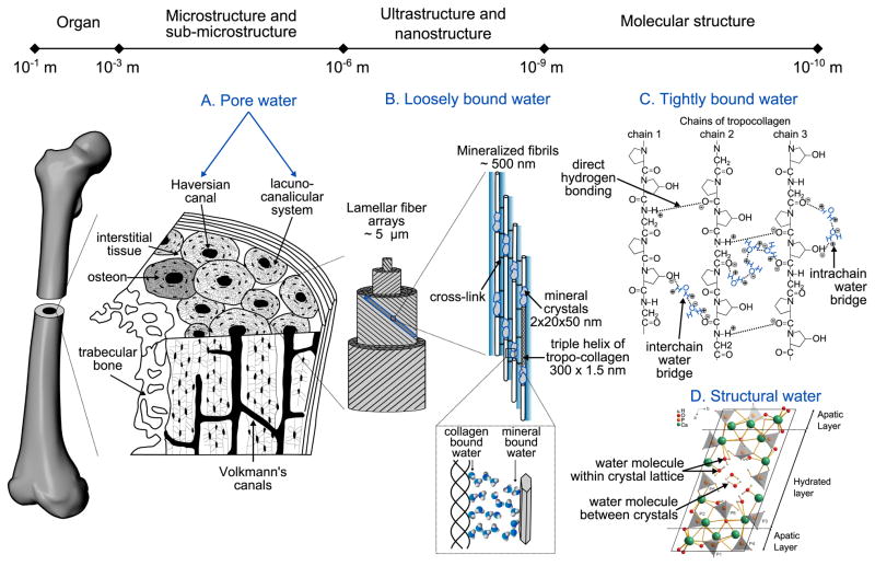
Fig. 1.
Schematic of the presence of water in bone at each hierarchical level of organization. Cortical tissue has a network of pores comprising vascular porosity (Haversian and Volkman's canals) and the lacuno-canalicular network. The bone matrix is arranged in arrays of lamellar fibers that are made up of collagen fibrils mineralized by apatite crystals. In this highly organized structure, water exists at different energy levels: free as liquid, loosely bound, tightly bound, and part of the mineral lattice. (A) At the microscale, free (or pore) water occupies the vascular-lacunar-canalicular space. (B) Loosely bound water is found at the surface of the collagen fibrils and between the collagen and the mineral phase. (C) At the molecular scale, tightly bound water refers to water molecules trapped inside the collagen triple helix. Examples of water bridges within a chain or between the chains of a tropocollagen molecule are illustrated on a small sequence of collagen (GLY-PRO-HYP) where hydrogen bonds are represented with dotted lines, and plus and minus signs indicate the polarity of the bonded atoms (adapted from Bella et al. [17] and Brodsky et al. [16]). (D) Structural water refers to the water molecules found within the core of the apatite structure (reprinted with the permission of PNAS from Davies et al. [28]).

