
Figure 4.
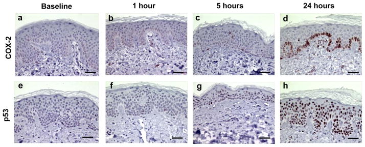
Examples of immunohistochemically stained proteins in sun-protected skin after 2–3 MED of SSL-irradiation. COX-2 expression at baseline (a), 1 hour (b), 5 hours (c), and 24 hours (d). p53 expression at baseline (e), 1 hour (f), 5 hours (g), and 24 hours (h). Images are at a magnification of 400X and the scale bar represents 50 μm. Brown nuclear and cytoplasmic stains represent positive and blue stain represents negative staining.