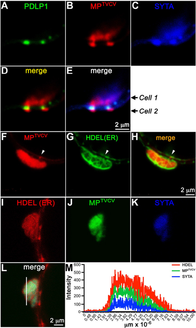
Figure 4. SYTA and MPTVCV co-localize in virus replication sites at PD in N. benthamiana.
(A–E) Confocal images of SYTA-CFP and PDLP1-GFP (PD marker) at late stages in a replicon TVCV::MP-RFP infected cell (Cell 1) in N. benthamiana show (A–C, E) SYTA and MPTVCV co-localizing in virus replication sites formed in the cell cortex adjacent to two PD, and (C, E) SYTA also accumulating within the PD, while (B, D) MPTVCV has moved into a neighboring cell (Cell 2).
(F–H) Confocal images of GFP-HDEL at late stages in a replicon TVCV::MP-RFP infected cell in N. benthamiana show MPTVCV accumulating in ER membrane-containing (HDEL marker) replication sites formed in the cell cortex adjacent to PD. MPTVCV, but not HDEL (ER), can also be seen within PD at some sites (arrow).
(I–L) Confocal images of SYTA-CFP and RFP-HDEL at late stages in a replicon TVCV::MP-GFP infected cell in N. benthamiana show SYTA, MPTVCV and HDEL (ER marker) co-localizing within a TVCV cortical replication site.
(M) Fluorescence intensities of SYTA-CFP, MPTVCV-GFP and RFP-HDEL across a section in the replication site (line in L) show MPTVCV and SYTA within the site caged by ER membrane.
Scale bars as indicated.

